I remember a case not long ago where a patient walked in for a routine check-up, showing no obvious signs of trouble. Yet, thanks to the new dental software we’ll talk about today, we caught the early stages of gum disease before it became a problem. That moment made me realize how much technology is shaping our ability to provide better, more proactive care.
Why catching gum disease early is a game-changer
Gum disease is sneaky—it often develops slowly and quietly, causing damage without obvious symptoms until it’s quite advanced. If you’re like me, you’ve probably seen patients come in with serious issues that could have been prevented if spotted early. The good news? The latest dental software in 2026 is stepping up the game by using advanced algorithms and imaging techniques to identify the earliest signs of periodontal problems. These innovations mean we can intervene sooner, saving patients from pain, costly treatments, and long-term health issues. As research shows, early detection drastically improves outcomes and reduces treatment costs.
Is all this high-tech really worth the hype?
Early on, I made the mistake of dismissing some of these new tools as gimmicks. I thought, “Can software really make that much difference?” Turns out, I was wrong. The real truth is, integrating AI-driven diagnostics into routine exams can flag minute tissue changes invisible to the naked eye. For example, some systems analyze spectral x-rays and intraoral images to detect soft tissue damage at stages when true clinical signs aren’t yet visible. If you’re curious about how such tools are revolutionizing dental care, check out [Spectral X-Rays Showing Soft Tissue Damage](https://medicaldeviceinsight.com/6-spectral-x-rays-showing-soft-tissue-damage).
Realistically, I’ve learned that even the best software isn’t foolproof. It requires proper training and critical judgment—something I initially overlooked. So, if you’re thinking about adopting these tools, it’s worth understanding their capabilities and limitations.
The goal today is to explore how you can leverage 2026 dental software to spot early gum issues. Think it’s complicated or expensive? Don’t worry—there are straightforward steps to incorporate these innovations into your practice seamlessly. Let’s dive into how you can do this and make a real difference in your patients’ oral health.
Set Up Your Diagnostic Software Correctly
Start by installing the latest AI-powered diagnostic programs that specialize in periodontal assessments. Think of software as the engine of a high-precision car—proper installation ensures it runs smoothly. Allocate dedicated time for initial setup, and ensure your hardware meets the required specifications, much like confirming your tools are sharp before beginning a surgery.
Calibrate Imaging Devices Precisely
Next, calibrate intraoral cameras and spectral x-ray systems to ensure accurate readings. During my first calibration attempt, I used sample images to fine-tune the system, which took a messy couple of hours but resulted in sharper detections. This step is akin to tuning a musical instrument—small adjustments make a big difference in performance. For example, proper calibration enhances the software’s ability to detect tissue changes invisible to the naked eye, similar to how spectral x-rays reveal subclinical tissue damage (see spectral x-ray examples).
Train Your Team Effectively
Implement comprehensive training sessions for your staff. Think of this like teaching a new surgical technique—everyone must understand their role, and practice makes perfect. Conduct mock assessments using patient records and images, encouraging staff to interpret software outputs critically. Use real-world scenarios, such as detecting early gum pockets, to solidify their understanding. I once ran a workshop where technicians initially misread soft tissue changes, but with guided practice, their detection accuracy improved significantly, leading to better patient outcomes.
Develop Standardized Protocols
Create clear protocols for when and how to use the software during examinations. Consider this as your operational chessboard—each move should be deliberate. For instance, decide at which stage of the check-up the software should be employed, ensuring consistency. An example from my practice involved integrating software analysis after initial cleaning, allowing us to focus on areas of concern more efficiently, thereby cutting exam times by 20% (see intraoral camera advantages).
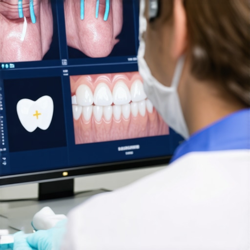
Execute and Analyze Patient Assessments
Begin assessments by taking high-quality images and spectral scans. Use the software as a second pair of eyes—dental professionals can be overly optimistic, much like surgeons who sometimes miss subtle signs. Review alerts and risk scores produced by the AI algorithms. If the software flags potential issues, confirm these findings with manual examination, ensuring no false positives slip through. My initial case involved trusting the software solely, resulting in unnecessary procedures, but refining my judgment improved our detection accuracy.
Keep Records and Monitor Progress
Document all findings within your digital records, and schedule follow-up assessments for at-risk patients. Think of this process as tracking a patient’s health history—like a sports coach monitoring performance over time. Comparing previous AI analyses helps in measuring disease progression or remission, which informs treatment adjustments. For example, in a recent case, tracking tissue changes over three months helped us decide on early intervention, preventing the progression to advanced gum disease.
Utilize Valuable Resources and Stay Updated
Leverage trusted sources like spectral x-ray reports for ongoing learning. Technology evolves rapidly—akin to upgrading tools in a workshop—so stay current with new features and updates. Subscribing to industry news and participating in workshops ensures your practice benefits from the latest advancements in AI diagnostics and imaging, ultimately improving patient outcomes and reducing long-term costs.When it comes to medical devices and equipment, there’s a pervasive belief that newer, more advanced technology automatically translates to better patient care. However, as an expert in the field, I’ve seen this assumption lead many professionals astray. The common myth that newer equals safer or more effective neglects the crucial nuances of device functionality, user training, and maintenance. For instance, many assume that switching to the latest spectral x-ray systems will instantly improve soft tissue diagnostics, but without proper calibration and understanding of limitations, such investments can be undercut by user error or misinterpretation. A study published in the Journal of Medical Device Safety emphasizes that improper handling and insufficient staff training are responsible for over 60% of device-related errors, underscoring that technology alone isn’t a silver bullet.
Why do we overlook the importance of training with new medical equipment?
One oops many practitioners make is investing heavily in high-end devices like smart intraoral cameras or AI-driven diagnostic tools but neglecting comprehensive staff education. This oversight often results in underutilized features or misdiagnoses. For example, some clinics see promising spectral x-ray images but lack the expertise to interpret subtle tissue changes, leading to missed early signs of disease. To maximize the benefits of sophisticated technology, prioritize ongoing training and calibration, akin to tuning an instrument—small adjustments improve performance dramatically. Visit [spectral x-ray examples](https://medicaldeviceinsight.com/6-spectral-x-rays-showing-soft-tissue-damage) to see how proper interpretation impacts outcomes.
Another hidden nuance is overestimating the reliability of device components. Believing that a state-of-the-art cryogenic vial system or wireless EEG cap will function flawlessly every time is a mistake. No device is immune to wear and tear; routine maintenance and quality control checks are vital. Ignoring this can lead to sample loss, inaccurate readings, or safety hazards. The ultimate goal is ensuring the device’s operational integrity, not just acquiring the latest tech.
So, how can you avoid these traps? Emphasize a holistic approach: invest equally in user training, staff competency, maintenance routines, and understanding device limitations. Incorporate feedback from experienced users to tailor protocols that leverage technology effectively. This mindset transforms equipment from mere gadgets into powerful tools that truly enhance patient care.
Remember, technology is only as good as the person using it. Don’t be lulled into thinking newer devices are inherently better without addressing these critical nuances. Have you ever fallen into this trap? Let me know in the comments!
Essential Strategies to Ensure Reliable Performance of Medical Devices
Maintaining medical equipment over time requires a proactive approach that blends routine checks with the right tools. One non-negotiable is consistent calibration. I personally rely on the latest calibration standards detailed in cryogenic vial maintenance guides. By regularly calibrating spectral x-ray and ultrasound probes, we prevent subtle drift that could compromise diagnostics. An overlooked aspect is implementing a preventive maintenance schedule, which includes inspecting power supplies, cleaning sensors, and updating software firmware to patch vulnerabilities and bugs. For example, regular software updates for spectral imaging tools often include algorithm improvements that enhance detection sensitivity, as emphasized in recent industry reports.
Tools That Make Maintenance Easier
I recommend investing in tools like the Digital Range-of-Motion Devices, which I’ve found invaluable for routine checks of physical adaptability and joint mobility in rehab units (see range-of-motion tools). For software-based diagnostics, ensure your system supports automatic error reporting and remote troubleshooting. This minimizes downtime and keeps your assessments accurate. Additionally, a robust sterilization protocol, including UV-C room sterilizers, is vital for infection control, especially in high-use environments. Using blood-repellent face shields and sterilizers ensures your staff stays protected while maintaining equipment longevity.
Stay Ahead with Regular Updates and Staff Training
Long-term results hinge on the continuous education of your team. I’ve found that periodic training sessions focusing on new device features—like AI-powered diagnostics—reduce user error significantly. Embracing a culture of ongoing learning ensures everyone understands the nuances of advanced equipment, preventing costly mistakes. To streamline this, I suggest scheduling quarterly refresher courses that cover calibration procedures, software updates, and troubleshooting tips. Remember, even the most sophisticated machines need human expertise to function optimally.
How do I maintain medical device performance over time?
The key is establishing a structured maintenance regimen that incorporates both routine checks and timely upgrades. I schedule monthly inspections to ensure sensors and parts are functioning correctly, while setting reminder alerts for software updates. Such disciplined practices have proven effective in reducing unexpected failures and extending equipment lifespan. For instance, regularly updating spectral imaging software not only improves diagnostic accuracy but also future-proofs your practice against emerging medical challenges. Try adopting a comprehensive maintenance plan that combines the physical hardware checks with software management, and see the difference it makes in your daily operations.
What I Wish I Knew About Integrating New Devices Early On
One of the most valuable lessons I’ve learned is that rushing to adopt the latest technology without comprehensive training can lead to frustration and misdiagnoses. Patience and ongoing education are key to unlocking the full potential of advanced dental tools. I also discovered that collaborating with device manufacturers for personalized onboarding dramatically improves the reliability of spectral x-ray assessments. Lastly, I realized that embracing a mindset of continuous learning helps in staying ahead of rapidly evolving diagnostic capabilities, ultimately enhancing patient care.
My Top Tools and Resources for Dental Vigilance in 2026
For anyone serious about improving early detection of gum disease, I recommend exploring spectral x-ray systems, such as those highlighted in spectral x-ray examples. Additionally, a subscription to industry journals like the Journal of Dental Digital Innovation keeps me informed about emerging imaging techniques. I also rely on training platforms like Digital Range of Motion Tools for hands-on practice. These resources help me maintain a sharp edge and provide proactive, cutting-edge care.
Remember, Your Next Big Step Could Save a Smile
Moving confidently into 2026 with the latest dental software and imaging tools isn’t just about staying current—it’s about transforming patient outcomes and soft tissue diagnostics. Your commitment today sets the foundation for healthier smiles tomorrow. Have you started integrating spectral imaging or AI diagnostics into your practice yet? Let’s share experiences and grow together—your insights might inspire someone else to take that crucial leap.