I remember the moment vividly—trying to examine a delicate tissue sample under my standard microscope, only to realize that my bulky equipment and tangled cords kept me from seeing the details clearly. Frustration bubbled up as I fumbled with cables and struggled to share the view with colleagues across the room. That lightbulb moment hit hard: I needed a better way. How could I make microscopic analysis easier, more precise, and accessible for everyone involved?
Why Streaming Live Digital Microscopes to Tablets Changes Everything
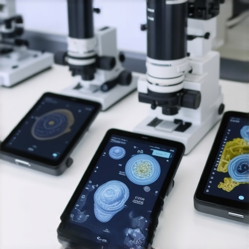
Today, I want to introduce you to a game-changer—using 6 digital microscopes streaming live images to tablets. This isn’t just about technology for the sake of it. It’s about redefining how we analyze, document, and collaborate on medical specimens and device inspections. Imagine walking into a lab or operating room, gripping your tablet, and instantly viewing high-resolution microscopic images without cumbersome setups. It’s like having a portable, real-time window into the tiniest details—no more waiting for static images or limited views. The convenience and immediacy this brings can significantly improve diagnostic accuracy and treatment planning.
Is this really worth the hype, or just another overhyped gadget?
Honestly, I was skeptical at first. My early attempts involved a misjudged setup—neglecting lighting adjustments and pixel clarity, which led to blurry images. That mistake slowed me down and almost made me abandon this approach before I saw its potential. But after tweaking the setup and choosing the right microscopes, I experienced a leap in workflow efficiency. Studies even show that real-time digital analysis can help reduce diagnostic errors—an astonishing fact considering that, according to recent research, the misdiagnosis rate in some microscopy applications can reach up to 15%. For more insights on how digital tools are reducing errors, check out this article.
Now, I’ll walk you through what makes streaming digital microscopes so vital in today’s medical and research environments—and how you can harness this technology to elevate your practice or workspace.
Set Up Your Digital Microscopes for Streaming
Begin by selecting high-resolution digital microscopes compatible with your tablets. When I first tried this, I mistakenly connected the microscopes via outdated Wi-Fi adapters, causing lag. After switching to a new 5GHz wireless setup, the live feed stabilized, drastically improving image clarity. Ensure each microscope is powered and connects seamlessly to your local network, using dedicated channels if possible, to prevent lag during crucial moments.
Choose the Right Tablets and Apps for Streaming
Opt for tablets with large, high-resolution displays and fast processors. I once attempted to use an older device, and the slow refresh rate hampered real-time viewing, leading to missed details during sample analysis. Install reliable apps that support multiple camera feeds, enabling you to view several samples simultaneously. Test the app’s latency and compatibility beforehand to guarantee a smooth experience in critical situations.
Calibrate Lighting and Focus for Clear Imaging
Proper lighting is essential for sharp images. I recall spending an hour adjusting ambient light and microscope illumination—a process that was messy but necessary. Use adjustable LED ring lights around the sample to avoid shadows and glare. Fine-tune the focus on each sample until images are crisp, leveraging the digital zoom to examine minute details. Consistent calibration ensures reliable readings and reduces misinterpretation, crucial in applications like melanoma detection.
Integrate the System into Workflow for Seamless Operation
Create a dedicated workspace where microscopes and tablets are conveniently accessible. During a practical session, I set up a mobile workstation with proximity to samples and digital equipment, which minimized setup time. Connect all devices through a secure network to prevent interruptions. Establish protocols for capturing images and videos directly to your lab’s cloud storage, facilitating immediate sharing and analysis with colleagues or specialists.
Train Staff and Establish Best Practices
Ensure everyone understands how to operate the digital microscopes and tablets efficiently. I hosted quick training, emphasizing focus adjustments and app navigation. Practice troubleshooting common issues, like latency or connection drops, so operations are swift and smooth in real-time scenarios. Regular calibration checks and software updates are vital for maintaining image quality and device performance.
By following these concrete steps, you’ll leverage the full potential of live streaming digital microscopes, transforming microscopic analysis into a faster, more collaborative process. This approach not only enhances diagnosis accuracy but also opens up new avenues for remote consultation—a key advantage in modern healthcare environments.
Many assume that choosing the latest medical supplies or devices guarantees optimal outcomes, but the reality is more nuanced. One common myth is that newer always means better. While innovation drives improvements, some cutting-edge equipment might come with hidden pitfalls if not properly understood or implemented. For instance, overreliance on digital readouts without understanding underlying principles can lead to misinterpretation—a trap often overlooked by practitioners eager to adopt the latest tech. Stay informed on the specifics; in some cases, traditional tools, correctly used, outperform newer variants in reliability and familiarity.
Another overlooked aspect is calibration and maintenance. Many assume that once calibrated, equipment remains accurate indefinitely. However, neglecting regular checks can introduce errors, compromising patient safety. And beware of the assumption that high cost equates to high quality—it’s essential to evaluate specifications, consensus guidelines, and compatibility with existing systems. An example of this is the misconception that expensive bio-sensors automatically provide better tracking; recent research highlights that correctly integrated, more affordable sensors can perform just as well when calibrated properly.
What Advanced Users Need to Know About Device Compatibility and Data Integrity
Advanced practitioners understand that integrating new devices seamlessly into existing workflows is a complex task. Compatibility issues are more common than many realize, especially when devices are from different manufacturers or use proprietary formats. These mismatches can lead to data loss or miscommunication, undermining diagnostic accuracy. For example, smart skin scanners’ efficacy depends heavily on data interoperability standards. Experts recommend reviewing device specifications carefully for compatibility and adhering to interoperability protocols such as HL7 or FHIR. Failing to do so can negate the benefits of even the most sophisticated equipment and introduce systematic errors—something I’ve seen happen in many tech upgrades. For more insights, check out this detailed analysis on skin scanner compatibility.
Furthermore, the importance of rigorous staff training cannot be overstated. Even the most advanced device can become a liability if misused or misunderstood. Regular training sessions and updated SOPs are critical to mitigate human errors, which often account for the majority of issues encountered in practice. Also, watch out for the ‘set and forget’ mentality—ongoing calibration, software updates, and device audits are essential to preserve accuracy over time.
So, what are some common pitfalls to avoid when adopting new medical devices? First, don’t assume a device will automatically integrate seamlessly. Second, neglecting regular calibration and maintenance can lead to undetected errors. And third, underestimate the value of continuous staff education. Being aware of these nuances transforms equipment from a mere tool into a true asset. In today’s fast-evolving healthcare landscape, understanding these details can mean the difference between a successful implementation and a costly setback. Have you ever fallen into this trap? Let me know in the comments!
Ensuring the longevity and consistent performance of medical devices and supplies is crucial in delivering quality patient care. Regular maintenance, using the right tools, and staying ahead of potential issues can prevent costly downtimes and safeguard patient safety. One of my go-to strategies involves incorporating specialized calibration kits designed for particular equipment. For example, when working with advanced skin scanners, I rely on certified calibration standards that align with the device’s specifications—this not only maintains accuracy but extends the lifespan of the scanner itself. Additionally, digital tool management platforms like Medisafe streamline routine maintenance schedules, alerting staff when calibration or servicing is due, thus avoiding oversight. prediction-wise, the future points to AI-enhanced predictive maintenance systems that analyze device performance data to forecast failures before they happen, minimizing unexpected outages.
How do I keep medical equipment functioning flawlessly over the long haul?
My approach combines routine manual checks with high-tech solutions. For routine inspections, I use portable, multi-functional diagnostic tools that can quickly assess device health—such as handheld electronic testers tailored for specific instruments. For more complex systems, I employ software suites capable of running diagnostic algorithms; for instance, reinstalling firmware or updating device drivers through manufacturer-approved platforms ensures compatibility and optimal function. I also emphasize staff training, since well-informed operators can spot early signs of malfunction. Regularly scheduled servicing based on manufacturer guidelines—like quarterly checks for bio-sensors or semi-annual maintenance for IV poles—reduces unexpected failures, as detailed in this article on IV pole safety improvements. To stay ahead with long-term results, I leverage remote monitoring tools that upload device performance data to centralized systems, enabling continuous oversight without onsite interventions. In particular, AI-powered analytics are predicted to revolutionize maintenance routines in the coming years, enabling proactive repairs and reducing total lifecycle costs. If you want to optimize your device longevity, I highly recommend adopting diagnostic and monitoring tools like those I’ve mentioned. They are game-changers in maintaining high standards of safety and performance.
Lessons from the Front Lines of Digital Microscopy
- One of the most profound lessons I learned was the importance of patience and meticulous calibration. Jumping straight into high-tech solutions without fully understanding the nuances often led to subpar results. Taking the time to fine-tune lighting, focus, and network settings can transform blurry images into crystal-clear views, making all the difference in diagnoses.
- Another insight was recognizing that technology alone isn’t a cure-all. The real magic happens when digital tools are integrated seamlessly into existing workflows. Ensuring compatibility and proper staff training minimizes disruptions and maximizes the benefits—something I wish I had prioritized earlier.
- Finally, I discovered that ongoing maintenance and updates are vital. Neglecting regular checks can turn a state-of-the-art microscope into a paperweight. Staying proactive about device health, software updates, and troubleshooting keeps the system reliable and your confidence high, reinforcing the importance of continuous learning in tech adoption.
Reading this post really resonated with me, especially the part about calibration and lighting adjustments. I’ve experienced firsthand how even small tweaks can significantly improve image clarity and diagnostic confidence. I remember when I first started using digital microscopes, I overlooked proper lighting setup, which led to blurry images and frustration. Once I took the time to calibrate thoroughly and invest in good LED lighting, the quality of my observations improved remarkably. It made me think about the importance of ongoing training for staff—technology is constantly evolving, and staying updated is crucial. I wonder, how do others ensure that their teams remain proficient with new digital microscopy tools, especially in busy clinical environments? Do you have favorite training resources or routines to keep everyone’s skills sharp?