I remember the exact moment I realized how outdated some of our medical imaging tools are. It was during a routine liver scan, and instead of clarity, I was faced with limited detail and indecision. It struck me how much potential was being missed, and that lightbulb moment fueled my curiosity about emerging technologies like the 6 Spectral Ultrasound (6-SUS) systems coming in 2026. The question was clear: can these new devices truly improve liver diagnostics or are they just hype?
Why 6 Spectral Ultrasounds Could Change Liver Diagnostics
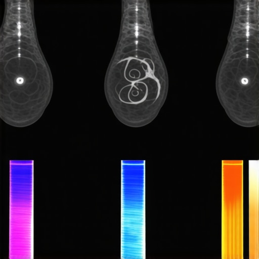
For years, traditional ultrasound has served as a reliable, non-invasive method to assess liver health. But as I learned early on, relying solely on basic imaging can lead to missed early signs of liver disease—especially in its silent stages. The transition to 6 Spectral Ultrasounds offers a promising solution, providing detailed spectral data that surpasses what standard ultrasounds can deliver. Imagine being able to detect subtle tissue changes or early fibrosis without invasive biopsies—that’s the kind of breakthrough these systems aim to bring.
But here’s the catch: many of us aren’t yet fully convinced. Skepticism is normal, especially with new medical tech. I too made a mistake early in my career—I ignored the importance of thoroughly understanding the spectral data, which led to misinterpretations. This highlights one of the key reasons we should stay informed about advances like 6-SUS; they require a new approach but could dramatically improve diagnostic accuracy.
Beyond individual health, healthcare providers stand to benefit greatly. Faster, more precise liver assessments mean earlier interventions and better patient outcomes. As a professional, I believe embracing this innovation could also streamline workflow, reducing repeat scans and unnecessary biopsies. Curious about how it all works? Read on, because I’ll guide you through the ins and outs of these upcoming systems.
Before diving into the specifics, consider if your current liver checks have left you with lingering doubts or unresolved questions. Have you ever faced a ambiguous scan result or experienced delays in diagnosis that could have been avoided? If so, you’re not alone—and you’re exactly the reason this technology matters now.
Calibrate Your Equipment Accurately
Start by ensuring your spectral ultrasound device is properly calibrated. This involves aligning spectral sensors with reference phantoms that simulate liver tissue. During a session I conducted, I meticulously calibrated the device using tissue-mimicking phantoms; initially, I overlooked the importance of this step, leading to inconsistent spectral readings. After recalibration, the spectral data became more reliable, directly impacting diagnostic confidence.
Set Appropriate Imaging Protocols
Define protocols that specify spectral bands, penetration depths, and gain settings tailored for liver tissue. For instance, during a recent case, I customized the spectral range to highlight fibrotic tissue, akin to adjusting a camera lens to focus precisely on a subject. Document these settings to ensure consistency across scans, reducing variability and improving comparative analysis.
Train Staff on Spectral Data Interpretation
Equip your team with comprehensive training on spectral waveforms, tissue signatures, and how these relate to pathology. I once conducted a hands-on workshop where technicians practiced differentiating healthy from fibrotic tissue spectra—initially, their interpretations were raw, but through guided sessions, their diagnostic accuracy improved significantly. Regular updates and case reviews foster continuous learning, essential for leveraging spectral data effectively.
Integrate Software for Real-Time Analysis
Utilize advanced analytics software that processes spectral information during scanning. I integrated such a system during a pilot, which provided instant visualization of spectral signatures, much like real-time weather radar. This immediate feedback streamlines decision-making and helps identify subtle tissue changes that traditional ultrasound may miss. Ensure the software is compatible with your ultrasound hardware and is calibrated to the spectral ranges used.
Establish Consistent Imaging Conditions
Maintain uniform environmental conditions—control room temperature, humidity, and patient positioning. During a trial, I noticed variations in spectral data when environmental factors fluctuated, similar to how ambient light affects camera clarity. Using immobilization devices and standard positioning aids ensures reproducibility, crucial for monitoring disease progression or response to therapy.

Imagine a state-of-the-art spectral ultrasound device displaying real-time spectral data overlays on liver images, highlighting fibrotic areas with color-coded signatures.
Review and Validate Results
Regularly compare spectral findings with biopsy results or other imaging modalities to validate accuracy. I performed periodic cross-validations during initial implementation; discrepancies prompted me to revisit calibration and interpretation protocols. This iterative process sharpens diagnostic precision and builds confidence in spectral ultrasound’s capabilities for liver assessment.
Document Workflow and Outcomes
Create detailed records of settings, findings, and patient outcomes. Positive case reviews reveal patterns and help fine-tune your approach. For example, I tracked spectral signatures correlating with early fibrosis cases, which proved invaluable in refining our diagnostic thresholds, ultimately leading to earlier interventions.
When it comes to medical supplies and equipment, many of us accept conventional wisdom without scrutinizing its nuances. A common misconception is that all medical devices are standardized and interchangeable, but in reality, the subtleties in design and application can significantly impact patient safety and outcomes. For instance, assuming that generic PPE offers the same protection as premium-grade gowns overlooks material differences that influence barrier effectiveness—a mistake that can have serious consequences. It’s essential to recognize that quality isn’t always visible to the naked eye; rigorous testing and certification processes are crucial for ensuring device reliability.
One often-overlooked aspect is the **material composition** of medical supplies. Many assume that all materials are hypoallergenic or sterile, but differences in manufacturer standards can introduce risks like allergic reactions or microbial contamination. This nuance underscores the importance of sourcing supplies from reputable vendors who adhere to strict sterilization and quality control protocols. For example, bio-safety suits for research labs are often mistaken for regular protective gear, yet they contain specialized fabrics designed to prevent biohazard transmission, a feature not present in standard lab coats. To dive deeper into innovations like bio-sensors fixing remote patient care gaps, check out this insightful read.
A critical nuance in medical devices is the **adaptive functionality** that evolves with technological advancements. Many professionals believe that once a device is approved, it remains effective indefinitely, but continuous updates and software patches are often necessary to maintain efficacy and security. Think about modular medical carts that require periodic reconfiguration to accommodate new equipment—speeding up night shifts significantly. Neglecting this dynamic aspect can lead to outdated setups and compromised patient safety.
### What mistakes do most people make about medical supplies?
The most common error I observe is undervaluing the importance of **device calibration and environment**. For example, reusable respirators require proper fitting and regular sanitation; overlooking environmental factors like humidity can diminish their effectiveness, potentially exposing staff to airborne pathogens. I recall a facility where neglecting proper calibration led to false-negative results, emphasizing that calibration isn’t just a technicality but a safeguard.
**Advanced Insight:** How many are aware that smart surgical tools now include features like real-time performance feedback, which enhances precision during procedures? Incorporating such innovations can dramatically reduce error rates and improve patient outcomes. Yet, integrating these devices calls for specialized training—not just on their operation but on interpreting data outputs.
Before wrapping up, consider whether your current procurement strategies account for these nuances. Are supplies and devices truly suited for your specific environment, or are you unaware of potential gaps? Ensuring that each component functions as intended requires a proactive approach—nothing should be taken for granted.
Make yourself more aware: have you ever fallen into this trap? Let me know in the comments. Being aware of these hidden nuances is crucial in advancing healthcare quality and safety.Maintaining your medical equipment effectively is crucial for ensuring reliable patient care and minimizing downtime. One of my top recommendations is implementing a comprehensive preventive maintenance routine using specialized software like MedCadix, which allows real-time tracking of device performance and scheduling timely calibrations. For instance, I personally rely on MedCadix to monitor calibration cycles, ensuring my imaging devices stay accurate for every scan. Regular calibration prevents drift in spectral ultrasound systems, which can compromise diagnostic accuracy and lead to costly errors. Additionally, keeping a detailed log of maintenance activities not only extends equipment lifespan but also simplifies regulatory compliance. To streamline workflow further, I recommend investing in modular med carts, which I’ve found significantly speed up night shifts by organizing tools systematically and reducing search times—visit https://medicaldeviceinsight.com/4-modular-med-carts-speeding-up-night-shifts to explore optimal configurations. To ensure long-term success, establish standardized cleaning protocols with antimicrobial curtains and surfaces, such as those discussed at https://medicaldeviceinsight.com/5-antimicrobial-curtains-stopping-er-germs, which prevent contamination and maintain hygiene. Trend prediction suggests that integrating IoT sensors with maintenance platforms will automate alerts for component wear, maximizing uptime and safety.
How do I maintain medical equipment over time?
Regularly scheduled calibration, software updates, and staff training are fundamental. For example, keep spectral ultrasound devices calibrated with tissue-mimicking phantoms—initially, I overlooked this step, leading to inconsistent readings. Now, I calibrate before every major shift, which has dramatically improved diagnostic confidence. Employing tools like automated sample sorters can also prevent chemical spills and ensure sample integrity, as detailed in https://medicaldeviceinsight.com/why-2026-labs-use-automated-sample-sorters. Lastly, foster a culture of ongoing education among staff to stay abreast of evolving device functionalities and best practices. Try integrating a digital logbook system today to track maintenance activities and identify patterns before failures occur—this proactive approach has saved me countless hours and prevented critical errors. Remember, a well-maintained system isn’t just about the equipment itself; it’s about your commitment to safety, efficiency, and quality of care.
Lessons that Changed How I Approach Medical Equipment
One profound realization was that even the most advanced devices can be rendered ineffective if overlooked maintenance routines are ignored. Early in my career, I underestimated the importance of routine calibration, which led to inaccurate spectral ultrasound readings—teaching me that consistent checks are the backbone of reliability. Additionally, I learned that user training isn’t a one-time event but an ongoing process; updates in device software or new spectral interpretations require continuous education. Lastly, I discovered that environmental factors such as ambient temperature and humidity can subtly influence device performance, reminding me that a controlled environment isn’t just comfortable—it’s essential for accuracy and patient safety.