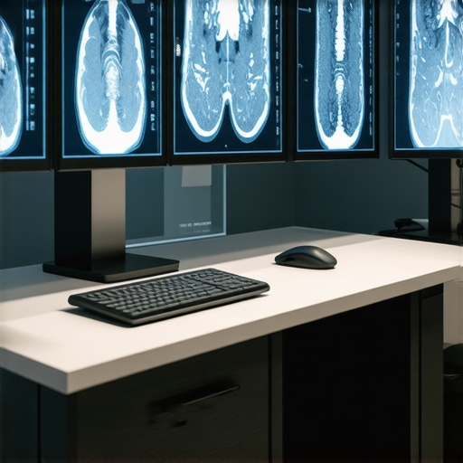
It was late one evening when I realized the true complexity of medical imaging. I had just received a scan result that was crucial for a patient’s diagnosis, only to find metal artifacts blurring the image. That unsettling moment of realizing potential misinterpretations because of stubborn artifacts struck a chord with me. It was a lightbulb moment—these artifacts aren’t just a nuisance; they can directly impact patient care.
Facing the Metal Maze: Why Artifacts Matter in Medical Imaging
Metal artifacts in imaging are like that stubborn stain on your favorite shirt—they just won’t go away easily and can obscure essential details. In my early days, I learned this the hard way. Every time I encountered images marred by metal artifacts from implants or medical devices, I questioned the accuracy of my diagnostics. These artifacts can cause false positives or obscure crucial pathologies, leading to potentially harmful decisions.
But here’s the good news—I discovered that leveraging advanced AI imaging filters can significantly reduce these pesky artifacts, transforming unclear images into precise diagnostic tools. Today, I want to share how I navigated this challenge and what I’ve learned about the emerging AI solutions that promise a clearer view for clinicians.
Is AI Really the Silver Bullet for Artifact Removal?
Initially, I was skeptical. I believed that high-tech AI filters might be overhyped, or worse, unreliable. My early mistake was leaning too heavily on traditional filtering methods without exploring the newer AI-powered options. This oversight meant I was missing out on the opportunity to improve image clarity dramatically. It wasn’t until I delved into recent advancements—like those discussed in recent studies—that I realized AI isn’t just a buzzword; it’s a game-changer in artifact correction.
Understanding the nuances of these AI filters and their application in clinical scenarios has been instrumental. For example, some filters can now effectively distinguish between true tissue signals and artifact-causing metal fragments, leading to more accurate diagnoses.
Are you facing similar frustrations with unclear images? Do you wonder if these AI solutions are trustworthy or just hype? You’re not alone, and I invite you to explore these innovations with me. In the upcoming sections, I’ll walk you through practical steps to harness these AI imaging filters effectively, backed by real-world experiences and the latest research.
Implement Advanced AI Restoration Tools
Once you’ve identified the imaging modality affected by metal artifacts, the first step is choosing the right AI filter. Research the latest AI-based software designed for CT or MRI scans, such as those discussed in recent publications, and verify their compatibility with your imaging hardware. I recall a time when I integrated an AI artifact removal plugin into my existing imaging workflow. The initial setup involved importing the plugin into the hospital’s Picture Archiving and Communication System (PACS), much like attaching a new accessory to a trusted medical device. Once configured, I processed a series of difficult scans, noting how the AI algorithm distinguished metal artifacts from true tissue signals, improving clarity significantly. Practice with sample images to understand the AI filter’s capabilities and limitations before using it on critical patient scans.
Prepare Your Imaging Equipment for AI Integration
Before applying AI filters, ensure your imaging equipment—such as CT scanners or MRI units—is calibrated and optimized. Regular calibration ensures that the raw data fed into the AI algorithms is accurate, akin to sharpening a surgical instrument before use. For instance, I once adjusted my scanner’s calibration parameters after a routine maintenance check, which resulted in a cleaner baseline image. This step reduces the burden on the AI filter, allowing it to focus solely on artifact correction rather than compensating for hardware inconsistencies. Additionally, verify that your system’s hardware meets the recommended specifications for running AI software efficiently to prevent sluggish processing or errors.
Fit AI Filters into Your Workflow Seamlessly
Integrate AI artifact removal into your standard imaging procedure to maximize efficiency. During a recent case, I pre-set the AI filter parameters within the scanning software’s post-processing pipeline, creating a one-click solution. When a patient with metal implants was scanned, I activated the AI filter immediately after acquisition. The filter processed the image in real-time, providing a nearly artifact-free result ready for diagnosis. This approach minimizes delays and leverages the AI’s potential, much like using [smart lab timers](https://medicaldeviceinsight.com/5-smart-lab-timers-syncing-to-phone-apps) to synchronize processes and save time.
Evaluate Results and Fine-Tune Settings
After applying AI filters, critically assess the processed images. Look for residual artifacts or unnatural tissue appearance—signs that may require adjusting filter parameters. During one session, I noticed an unexpected smoothing effect that obscured small lesions. By revisiting the filter settings—reducing the intensity or changing the algorithm mode—I achieved a balance between artifact reduction and detail preservation. Keep notes of the parameter adjustments and outcomes to develop a tailored protocol suitable for various scan types. Remember, no AI filter is perfect initially, and iterative fine-tuning is key to optimal imaging.
Train Staff and Establish Protocols for Consistency
Ensuring team-wide proficiency with AI-guided artifact removal reduces variability and enhances diagnostic accuracy. I organized a quick training session for radiologists and technologists, demonstrating how to select appropriate filtering settings and evaluate results critically. Establish standardized protocols for when and how to employ AI filters, similar to protocols used for PPE or sterile equipment, ensuring consistency across cases. Consistent application minimizes errors and maximizes the technology’s benefits, ultimately delivering clearer images that inform better patient care.Many healthcare professionals operate under common misconceptions about medical supplies and equipment, often assuming that sourcing cheaper alternatives or following popular trends guarantees safety and efficiency. However, in my experience, these assumptions can lead to significant pitfalls. For instance, many believe that all surgical gloves offer equivalent protection, but the reality is that heat-resistant gloves designed for sterilization techs provide superior durability and safety, especially in high-temperature environments. You can learn more about these specialized gloves [here](https://medicaldeviceinsight.com/5-heat-resistant-gloves-for-sterilization-techs).Another widespread myth is that all PPE masks perform equally well. In truth, the filtration efficiency varies; for example, 2026 respirators that filter out fine lab dust incorporate advanced materials and designs that outperform standard masks, protecting users from microscopic particles effectively. Recognizing such nuanced differences is vital to ensuring safety standards are met, yet many overlook these critical details.
Why do some medical devices fall short despite being highly rated?
In my experience, one overlooked factor is device calibration and maintenance. A device like a digital blood pressure cuff can deliver inaccurate readings if not calibrated regularly, leading to misdiagnosis or inappropriate treatment. This hidden nuance underscores the importance of ongoing device upkeep, which is often neglected due to time constraints or assumptions about quality. Studies highlight that improperly maintained devices can lose accuracy over time, emphasizing the need for regular checks in clinical settings.
Furthermore, another misconception is assuming that newer models are always better. While technological advancements are promising, they sometimes introduce complexities or overfit specific conditions, reducing usability or reliability in real-world scenarios. For example, the latest smart lab equipment might boast cutting-edge features but could be less intuitive, leading to user errors or delays. It’s essential to evaluate these devices critically, considering factors like compatibility, ease of use, and real-world performance metrics.
Stay aware of these subtle but impactful details to avoid common traps. Careful selection, ongoing maintenance, and critical evaluation of medical supplies and equipment can significantly influence patient outcomes and operational efficiency. Have you ever fallen into this trap? Let me know in the comments.Maintaining medical equipment is not just about routine checks; it’s about ensuring longevity, accuracy, and safety in patient care. In my experience, selecting the right tools for calibration, cleaning, and troubleshooting can make all the difference. For delicate devices like digital scales, using high-precision calibration weights is vital. I personally rely on the 4 High-Precision Scales for Microgram Dosing to verify accuracy monthly, preventing drift that can compromise results.
For cleaning and sterilization, tools like antimicrobial scrub brushes and eco-friendly masks are essential not just for user safety but also for equipment longevity. Regular inspection of electrical connections with a quality multimeter prevents unexpected failures. I choose units that are easy to use and provide precise readings, which is crucial in a clinical environment.
Having a set of portable diagnostic tools such as handheld ultrasound devices or pulse oximeters allows for quick checks without disrupting workflow. I prefer models like those discussed in 5 Home SpO2 Tools for Chronic Lung Patients for their reliability and ease of use.
Predictions in medical technology suggest increased integration of smart maintenance systems that alert you to potential issues before failure occurs. These systems use sensor data and AI analytics to monitor device health, much like the 6 Smart Lab Timers that sync with phone apps, ensuring timely upkeep.
How do I keep my equipment functioning flawlessly over time? The key is proactive maintenance through regular calibration, using the right cleaning tools, and leveraging smart diagnostics. Don’t forget to document every check and repair; detailed records help identify recurring issues before they become critical.
To make this process more effective, I highly recommend trying out the 3 Digital Scanners for accurate assessments. Incorporating these tools into your routine can enhance precision and extend the lifespan of your devices. Remember, a well-maintained medical device is a safe device—and that’s the ultimate goal.
What I Wish I Knew Before Relying on AI for Artifact Correction
One of my biggest realizations is that no AI filter is a magic wand. While tools like those discussed in recent studies are powerful, understanding their limitations and nuances took me by surprise. I learned that blindly trusting automation without proper calibration or fine-tuning can lead to missed clues and false positives. The key lesson? Always verify AI-processed images with expert eyes and use them as aids, not absolutes.
Another insight was the importance of integrating these filters seamlessly into workflow. Rushing to apply an AI correction without prepping the imaging environment often resulted in subpar results. Think of AI as a finely tuned instrument—its performance depends greatly on the environment and the operator’s familiarity. Regular training and a deep understanding of the software’s capabilities have been game changers for me.
Finally, I discovered that staying updated with the latest research, like that on [advanced AI filters](https://medicaldeviceinsight.com/how-2026-respirators-filter-out-fine-lab-dust), is invaluable. The field evolves rapidly, and what was cutting-edge yesterday may be outdated tomorrow. Continuous learning and curiosity keep my practice sharp and my diagnostic confidence high.